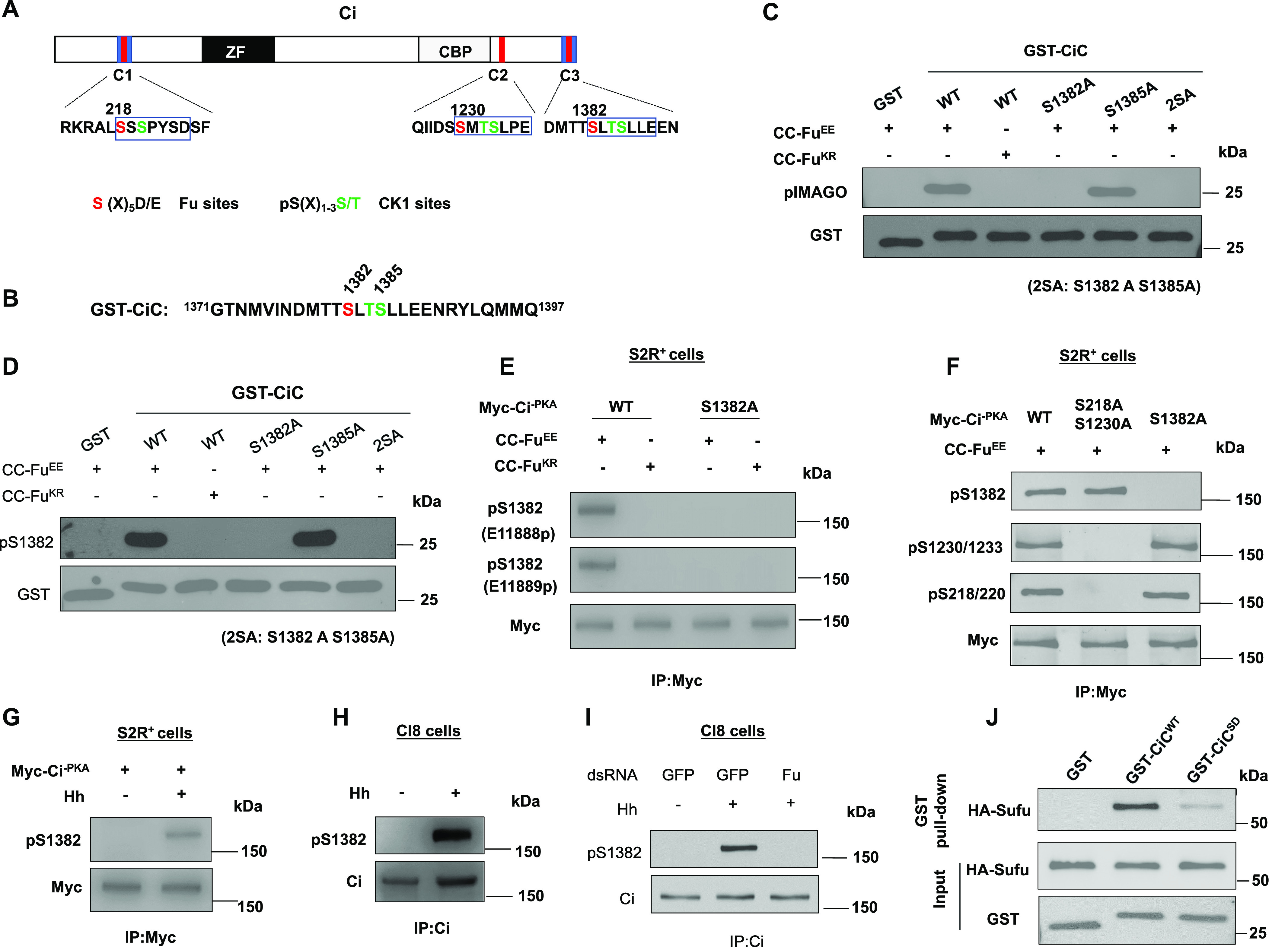
Figure 1. Fu phosphorylates the C-terminal Sufu-binding domain of Ci.

(A) Schematic diagram of Ci. Blue boxes and red bars indicate the Sufu-binding domains and Fu/CK1 phosphorylation clusters, respectively. ZF, Zinc-Finger DNA-binding domain; CBP, CBP-binding domain. The primary sequences of three phosphorylation clusters are shown with Fu and CK1 sites color-coded in red and green, respectively. (B) The primary sequence of the Ci C-terminal region in GST-CiC. (C, D) In vitro kinase assay using purified CC-FuEE or CC-FuKR as kinase and the indicated GST-CiC fusion proteins as substrates. Phosphorylation was detected by pIMAGO (C) or pS1382 antibody (D). (E) Western blot analysis of Ci phosphorylation in S2R+ cells transfected with the indicated Ci and Fu constructs. Two phospho-specific antibodies (E11888p and E11889p) were used to detect S1382 phosphorylation. (F) Western blot analysis of Ci phosphorylation on the indicated sites in S2R+ cells transfected with CC-FuEE and the indicated Myc-Ci-PKA constructs. (G) Western blot analysis of Ci phosphorylation on S1382 in S2R+ cells transfected with Myc-Ci-PKA and treated with or without Hh-conditioned medium. (H) Western blot analysis of endogenous Ci phosphorylation on S1382 in Cl8 cells treated with or without Hh-conditioned medium. (I) Western blot analysis of endogenous Ci phosphorylation on S1382 in Cl8 cells treated with the indicated dsRNA in the absence or presence of Hh. (J) Western blot analysis of HA-Sufu pulled down by the indicated GST fusion proteins. 5 μg of GST or GST-CiC fusion proteins were incubated with equal amounts of cell lysates from S2R+ cells transfected with HA-Sufu construct.
