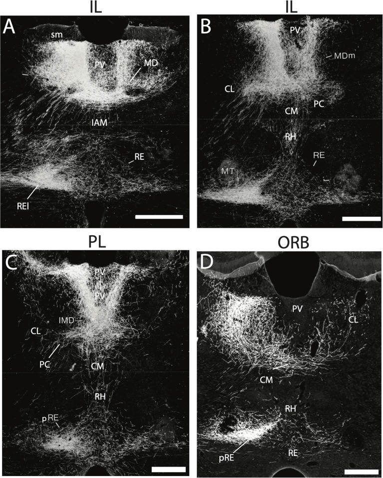
Figure 2.
(A–D) Darkfield micrographs of transverse sections through the thalamus depicting patterns of anterograde labeling produced by PHA-L injection in the infralimbic (IL) (A,B) and prelimbic (PL) (C) cortices of the medial prefrontal cortex (mPFC) and the ventral orbital cortex (ORB) (D). As depicted, injections in IL (A,B) and PL (C) gave rise dense terminal labeling of the paraventricular nucleus (PV) and medial division of mediodorsal nucleus (MDm), dorsally and rhomboid (RH) and the nucleus reuniens (RE), ventrally, with intense labeling of the lateral wings of RE (REl), rostrally (A) and peri-reuniens (pRE), caudally (B,C). By comparison, injections in the ventral orbital cortex (ORB) (D) produced dense labeling of the central medial (CM), paracentral (PC) and central lateral (CL) nuclei of the rostral intralaminar complex, heaviest in CL, ipsilaterally (left side) as well as pronounced labeling of the nucleus reuniens – comparable to that seen with injections in IL and PL (A–C). IAM, interanteromedial dorsal nucleus of thalamus; IMD, interomediodorsal nucleus of thalamus; MD, mediodorsal nucleus of thalamus; MT, mammillothalamic tract; sm, stria medullaris. Scale bar for (A–D) = 450 μm. Figure modified from Vertes (2002) and Hoover and Vertes (2011).

