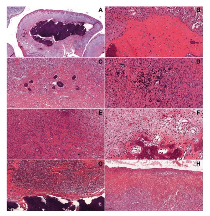
Figure 2.
Histopathological features of the peripheral ossifying fibroma (hematoxylin and eosin) - Lesions showing deposition of (A - 1000 µm) mature bone, (B - 100 µm) immature bone, (C - 200 µm) cementum-like tissue, and (D - 50 µm) dystrophic calcification. Bone tissue deposition in (E - 100 µm) hypercellularized and (F - 50 µm) hypocellularized areas. Chronic inflammatory infiltrate close to mineralized tissue (G - 200 µm). Ulceration (H - 200 µm).

