
Figure 1.
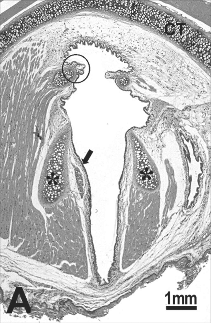
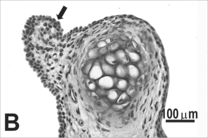
Control group: A: overview of the larynx demonstrating the thyroid (CT) and arytenoid (asterisk) cartilages, muscles, connective tissue, seromucous gland, free edge of the vocal fold (circle) and middle portion of the vocal fold (arrow); B: cuboidal epithelium (arrow) of the free edge of the vocal fold; C: stratified squamous epithelium (arrow) of the middle portion of the vocal fold. H.E.