
Figure 5.
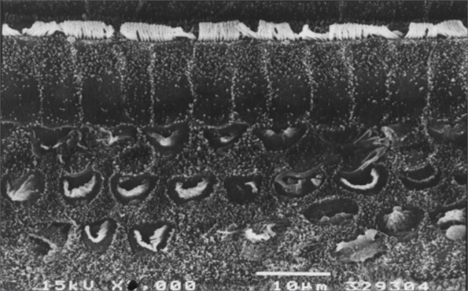
Scanning electron microscopy of a group 5 animal (EA and cisplatin treated during 3 weeks). Note extensive OHC injury in the basal portion of the cochlea. Scale = 10µM; 2000X magnification.
Official websites use .gov
A
.gov website belongs to an official
government organization in the United States.
Secure .gov websites use HTTPS
A lock (
) or https:// means you've safely
connected to the .gov website. Share sensitive
information only on official, secure websites.
Scanning electron microscopy of a group 5 animal (EA and cisplatin treated during 3 weeks). Note extensive OHC injury in the basal portion of the cochlea. Scale = 10µM; 2000X magnification.