
Figure 1.
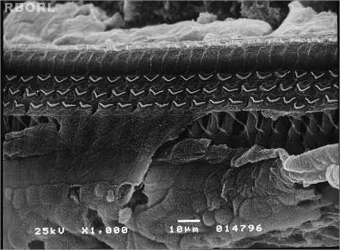
Photomicrography of a normal Corti's organ found in the guinea pig of group 1, showing its basal turn. Observe the uninjured structures in A: digital extension of the Deiter's cell; B: outer hair cells; C: outer hair cells estereocilia; D: reticular membrane. Magnified 1000x.