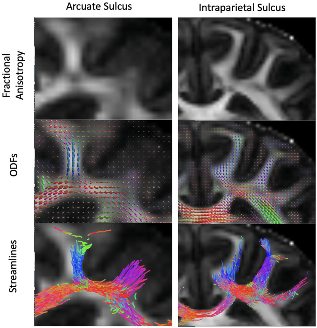
Fig. 10.
U-fibers. Fractional anisotropy maps for the arcuate and intraparietal sulci that contain short association fibers, also known as U-fibers. The middle panel shows the ODFs in the corresponding region. The lower panel displays streamlines generated from a seed that straddles the gray matter/white matter boundary as described by Reveley (Reveley et al., 2015a).

