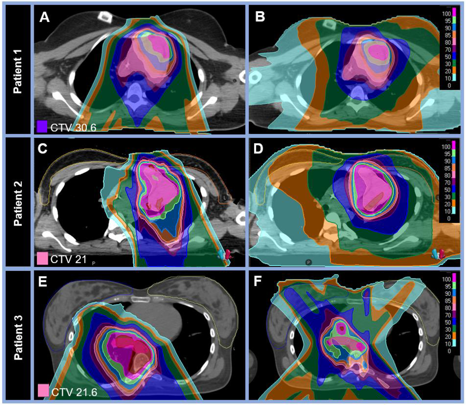
Figure 1: Proton therapy dose distributions with comparison photon plans for three female patients treated for Hodgkin lymphoma demonstrating reduced breast dose.

A-B: Comparative dose distributions on two axial planes for a (A) proton and (B) photon plan for an 18-year-old female patient with stage IIA HL of the mediastinum and left supraclavicular nodes treated with pencil-beam scanning protons to 30.6 Gy (RBE) in 17 fractions. Mean ipsilateral breast dose was 0.06 vs 1.53 Gy (RBE) and mean contralateral breast dose was 0.01 vs 0.99 Gy (RBE) for the proton vs photon plans, respectively. C-D: Comparative dose distributions on two axial planes for a (C) proton and (D) photon plan for a 16-year-old female patient with stage IVB HL treated with pencil-bream scanning protons to 21 Gy (RBE) in 14 fractions to the bilateral neck and mediastinum. Mean ipsilateral breast dose was 1.41 vs 2.85 Gy (RBE) and mean contralateral breast dose was 0.26 vs 1.23 Gy (RBE) for the proton vs photon plans, respectively. E-F: Comparative dose distributions on two axial planes for a (E) proton and (F) photon plan for a 21-year-old female patient with stage IVBX HL treated with pencil-bream scanning protons to 21.6 Gy (RBE) in 12 fractions to the mediastinum. Mean ipsilateral breast dose was 0.18 vs 5.62 Gy (RBE) and mean contralateral breast dose was 0.06 vs 3.50 Gy (RBE) for the proton vs photon plans, respectively. Detailed dosimetric data for other organs at risk are outlined in the supplemental Table.
