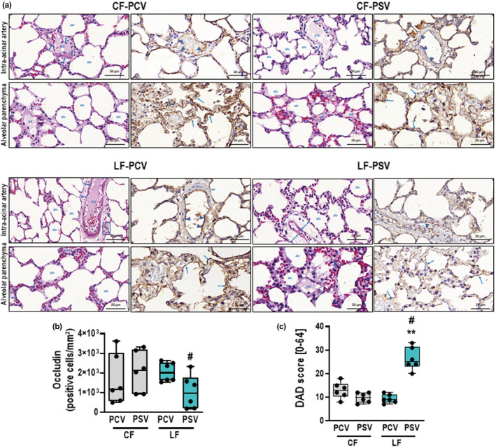
FIGURE 2.

DAD score and immunohistochemistry for occludin. (a) Representative images of histologic sections of alveolar pulmonary parenchyma (alveolar sacs and ducts, alveoli, and associated capillary loops) and perivascular sections (intra‐acinar arteries) stained by hematoxylin‐eosin and immunohistochemistry for occludin at high magnification. The intra‐acinar artery sections of CF‐PCV demonstrate discrete cuff‐shaped perivascular edema (double blue arrowheads) coincident with prominent expression of occludin by the endothelial cells of the artery (blue arrowheads). A similar immunophenotypic pattern of occludin was found in CF‐PSV and LF‐PCV, except for slightly cuff‐shaped perivascular edema (blue arrowheads) and mild inflammatory cells (blue square). In contrast, the LF‐PSV alveolar parenchyma exhibited almost absence of occludin in the endothelium of capillary loops (blue arrows). (b) Boxplot of occludin quantification in lung tissue. p = 0.026. (c) DAD score. **, means different from CF‐PSV (p < 0.01); #, vs LF‐PCV (p < 0.05). Alv, alveoli; art, artery; CF, conservative fluid therapy; DAD, diffuse alveolar damage; ed, edema; LF, liberal fluid therapy; PCV, pressure‐controlled ventilation; PSV, pressure‐support ventilation.
