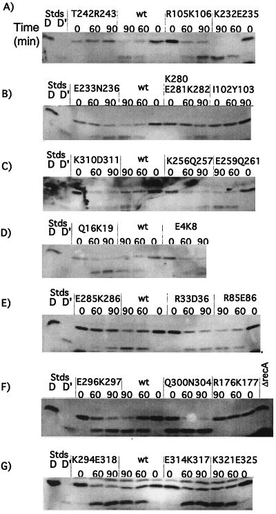
FIG. 4.
In vivo cleavage of UmuD by mutant RecA proteins. Derivatives of JL2460 carrying pTrecA220 or mutant derivatives were grown and treated as described in Materials and Methods; the strain marked ΔrecA carried pBR322. Each panel represents the results of a representative experiment. For each mutant strain, a time course is shown. Lanes marked “D” and “D′” are standards (Stds) from cells overproducing UmuD and UmuD′, respectively, and serve as markers. The lanes marked “0” contain samples taken without treatment; those marked “60” and “90” were taken 60 and 90 min after addition of nalidixic acid (50 μg/ml). The samples were analyzed by Western blotting with antibody to UmuD, as described in Materials and Methods. The name of the mutation is shown above each set of samples. wt, wild type.

