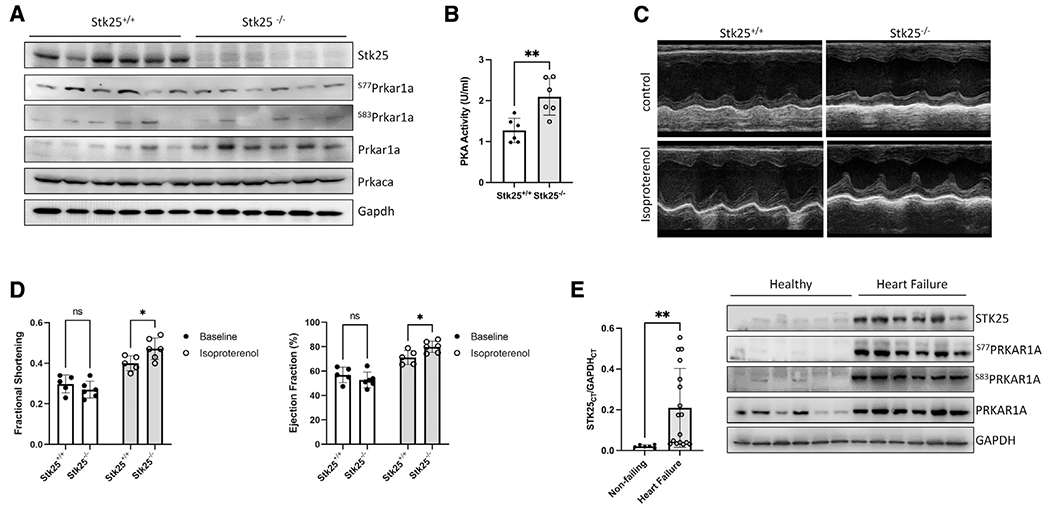
Figure 4. Stk25 loss increases response to adrenergic stimulation in vivo.

(A) Immmunoblot of Stk25, Prkaca, Gapdh, phospho-S77, phospho-S83, and total Prkar1a in Stk25+/+ and Stk25−/− whole-heart lysates.
(B) Stk25+/+ and Stk25−/− mouse heart lysates were assessed for PKA activity in vitro.
(C) Representative m-mode images of Stk25+/+ and Stk25−/− mouse hearts stimulated with either control or isoproterenol.
(D) Echocardiographic measurements of ejection fraction (EF) and fractional shortening (FS) at unstimulated baseline and in response to isoproterenol, n = 5 for Stk25+/+ and n = 6 for Stk25−/−.
(E) RT-PCR (left) from left ventricular myocardium of normal hearts (n = 6) or heart failure (n = 17) expressed as a ratio of the threshold cycle curve (Ct) of STK25 to GAPDH. Immunoblot (right) of STK25 and PRKAR1A expression and phosphorylation in protein lysates from left ventricular myocardium of normal hearts or failing hearts.
Bar graphs presented as mean ± SD and analyzed in technical triplicates, *p < 0.05, **p < 0.01 by Student’s t test in (B), repeated measures two-way ANOVA with Sidak’s correction for multiple comparisons in (D), and Welch’s t test in (E).
See also Figures S3 and S4 and Table S1.
