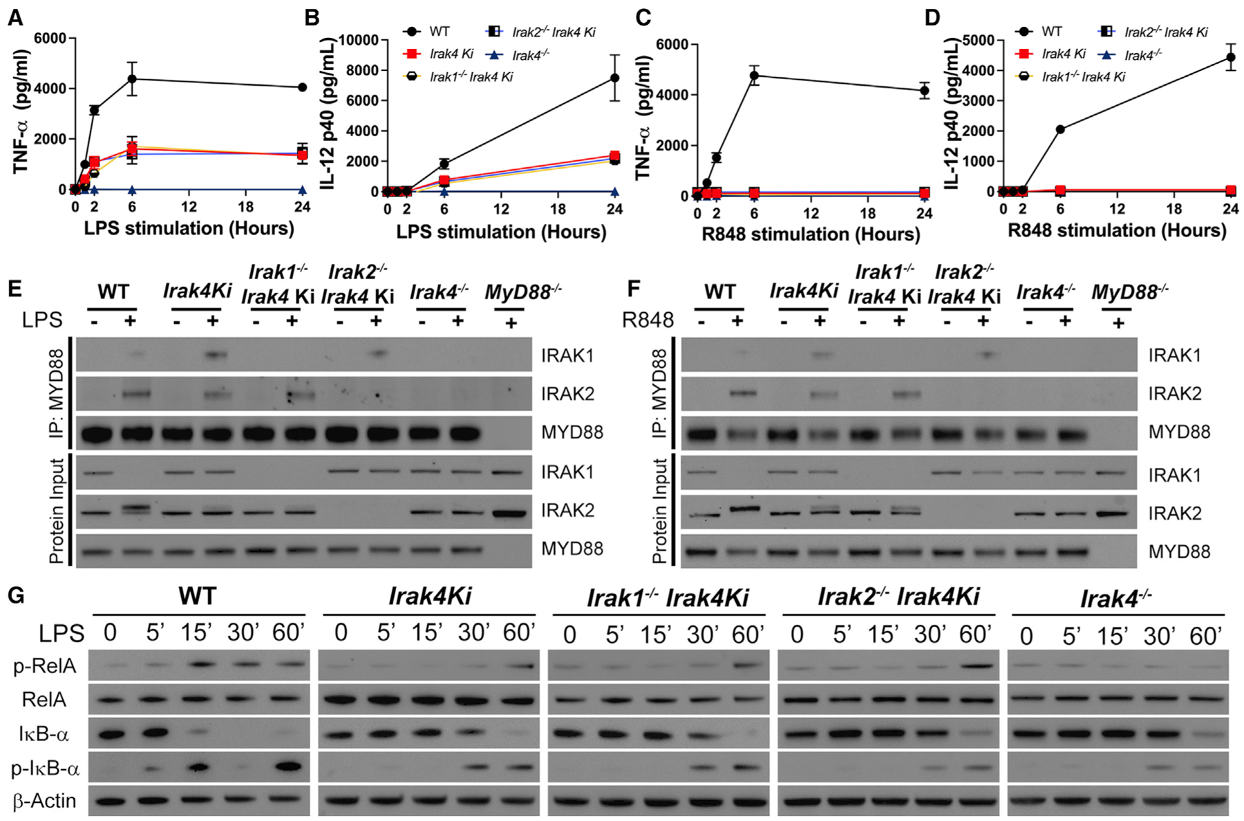
Figure 2. IRAK4 kinase activity is partially required for TLR4 signaling and essential for TLR7.

(A–D) Quantification of TNF-α (A, C) and IL-12 (B, D) produced after stimulation of WT, Irak4 Ki, Irak1−/−Irak4 Ki, Irak2−/−Irak4 Ki, and Irak4−/− BMDMs with LPS (A and B) or R848 (C and D) for up to 24 h.
(E and F) Immunoblot analysis of MYD88 co-immunoprecipitation with IRAK-1 and -2 in WT, Irak4 Ki, Irak1−/−Irak4 Ki, Irak2−/−Irak4 Ki, and Irak4−/− BMDMs treated for 60 min with LPS (E) or R848 (F).
(G) Kinetic study of p-RelA, RelA, IκB-α, p-IκB-α, and β-actin by immunoblot of whole cell lysates from indicated BMDMs treated with LPS for up to 60 min. All stimulations were done with LPS 100 ng mL−1 or R848 1 μg mL−1. *p < 0.05 in comparison with WT (one-way analysis of variance with Tukey’s multiple comparisons test). (A–D) Data from three independent experiments (mean and SEM). (E–G) Images are representative of three (E) or four (G) independent experiments.
