Fig. 3.
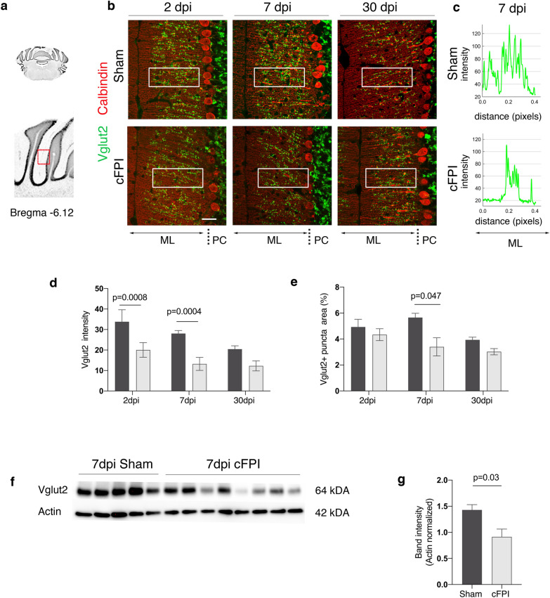
Decreased Vglut2 expression in the cerebellum of brain-injured mice at seven days after TBI. a Schematic of anterior coronal cerebellum sections (bregma -6.12). b Immunofluorescence labelling for Vglut2 (green) and calbindin (red) in the ML of the cerebellum of sham-injured and cFPI mice at 2, 7, and 30 dpi. c) Plots of signal fluorescence intensity profiles of Vglut2 expression from boxed areas in the cerebellar molecular layer (ML) of sham-injured and cFPI mice at 7dpi. d, e Quantification of Vglut2 density (d) and numbers of Vglut2 + puncta (e) from boxed areas in the ML at all time points after the injury. f, g Representative Western blots (f) and quantification (f, g) of Vglut2 band intensity in sham-injured and cFPI mice at 7 dpi (mean ± SEM; n = 5 (Sham), n = 8 (cFPI); two-tailed Student’s t-test, unpaired). PC Purkinje cells, dpi days post-injury, cFPI central fluid percussion injury, Vglut2 vesicular glutamate transporter-2

