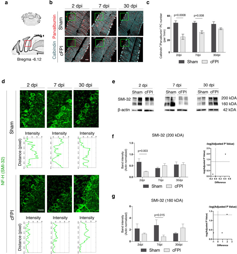
Fig. 4.
Reduced calbindin, parvalbumin, and non-phosphorylated neurofilament expression in the Purkinje cells after traumatic brain injury. a Schematic of anterior coronal cerebellum sections (bregma-6.12). b, c Representative confocal images (b) and quantification (c) of Purkinje cells (PCs) in the red boxed area in a co-labelled for calbindin (cyan) and parvalbumin (red) in sham-injured and cFPI mice at 2, 7, and 30 dpi, scale 50 μm. d High magnification confocal images and fluorescence intensity profiles of non-phosphorylated neurofilament-H (SMI-32) expression in Purkinje cells in the sham-injured and brain-injured mice at 2, 7 and 30 dpi in green boxed areas in b, scale 20 μm. e–g Representative Western blots (e) and quantification (f, g) of SMI-32 (200kDA) and SMI-32 (160kDA) band intensity in sham-injured and cFPI mice at 2, 7 and 30 dpi (mean ± SEM; n = 5 (Sham), n = 8 (cFPI); two-tailed Student’s t-test, unpaired). dpi days post-injury

