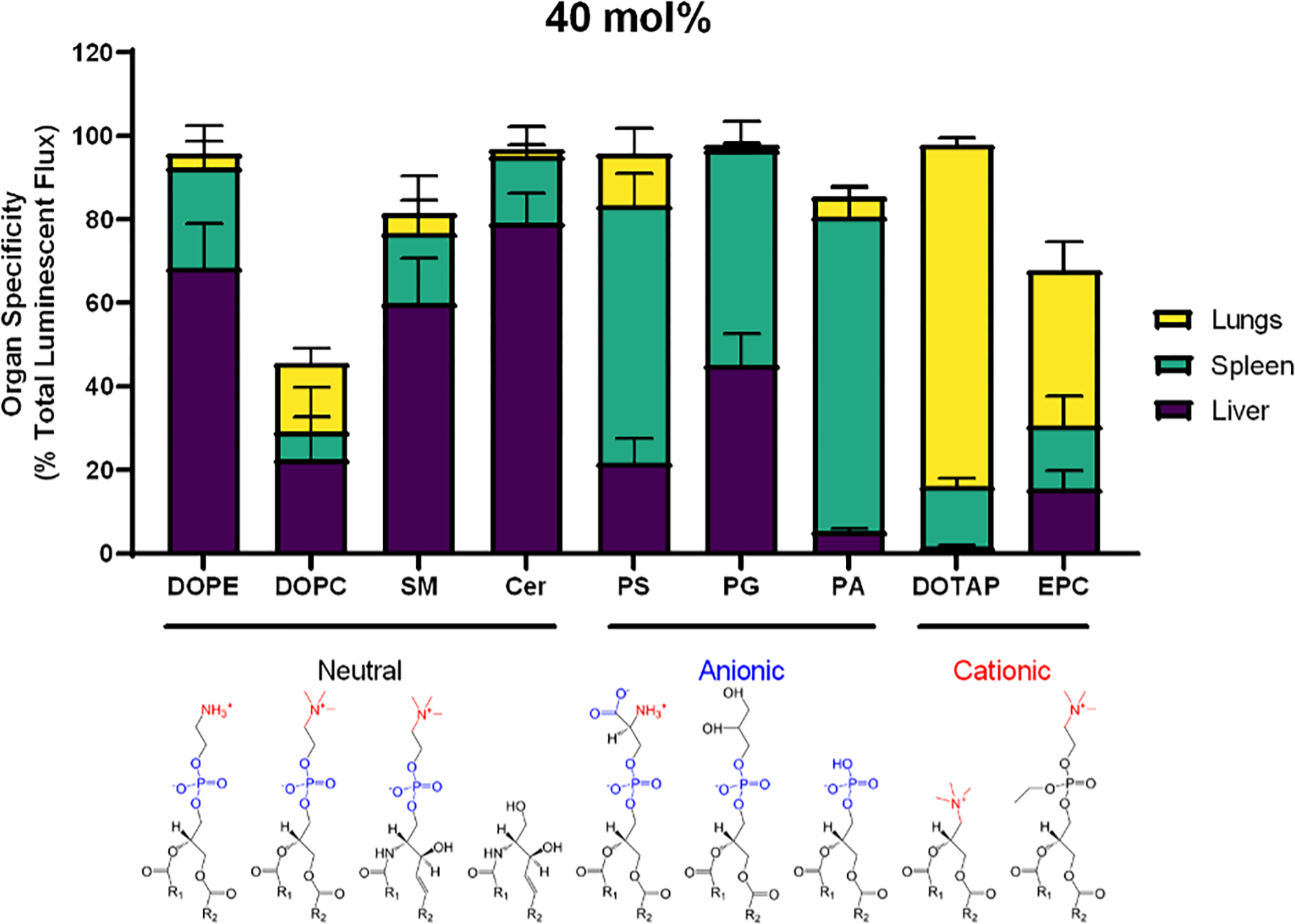
Figure 2: The effect of helper lipid charge on the location of protein expression holds is conserved across helper lipid chemistries.

LNPs were formulated with the ionizable lipidoid 306O10 and 40 mol% helper lipid and IV injected into mice (0.75 mg/kg mRNA). Luciferase signal was measured three hours later. In addition to DOPE, neutral lipids 1,2-dioleoyl-sn-glycero-3-phosphocholine (DOPC), sphingomyelin (SM), and ceramide (Cer) induced protein expression primarily in the liver. Anionic lipids, including PS, phosphatidylglycerol (PG) and phosphatidic acid (PA) shifted expression to the spleen, and the cationic lipids DOTAP and ethyl phosphatidylcholine (EPC) shifted expression to the lungs. Luminescence values as a percentage of total luminescence of all organs (liver, lungs, spleen, kidneys, intestines, pancreas, and heart) are plotted above. Mean values shown. Error bars represent standard deviation (n = 3).
