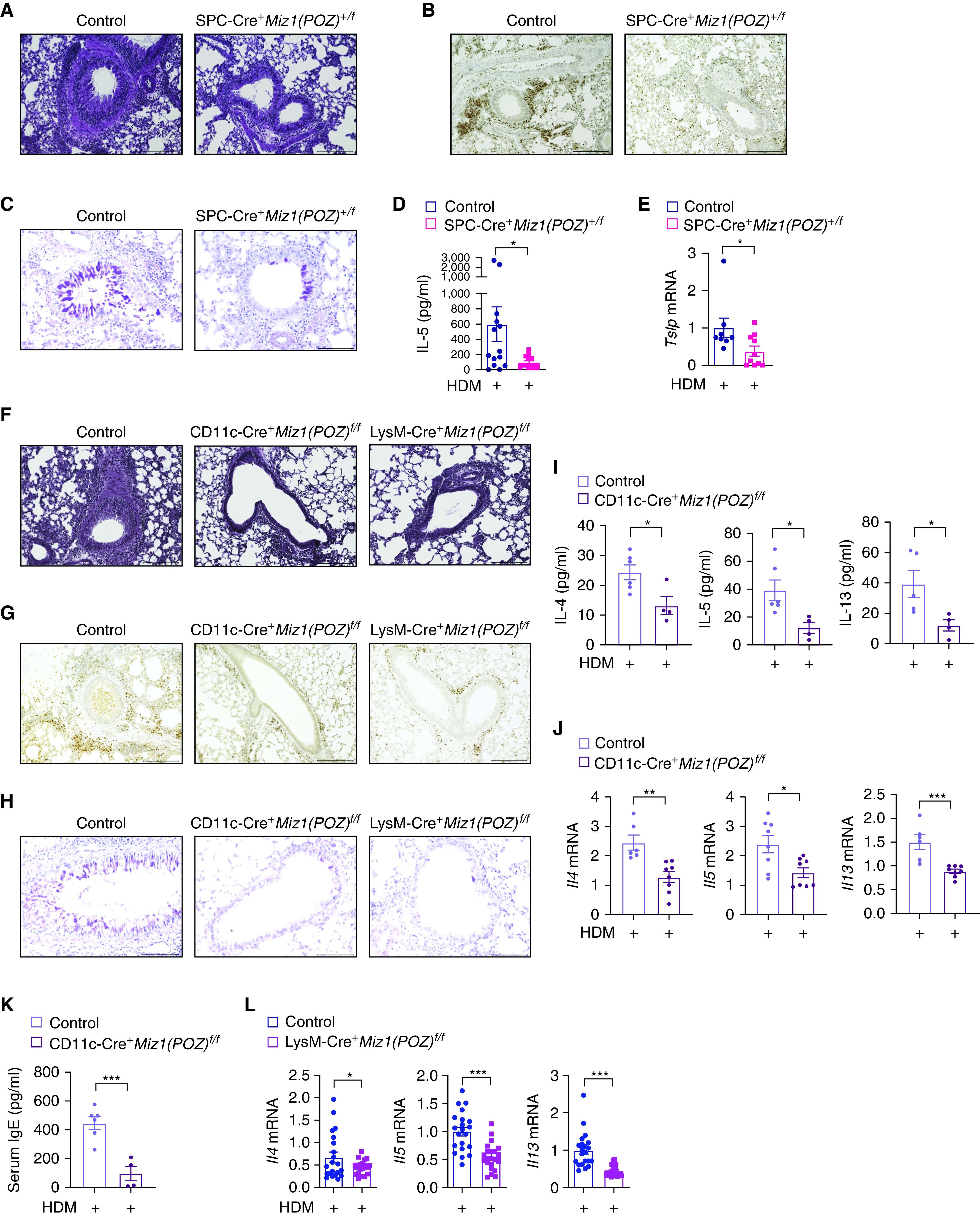
Figure 3.

Miz1 in lung epithelial cells or dendritic/myeloid cells contributes to HDM-induced allergic asthma. (A–C) Lung histology by hematoxylin and eosin staining (A), eosinophil major basic protein (EMBP) staining of eosinophils (B), or Periodic Acid–Schiff (PAS) staining (C) in the airways of HDM-treated control Miz1(POZ)fl/fl or heterozygous SPC-Cre+/Miz1(POZ)+/f mice. Eosinophils are stained brown/dark brown by EMBP staining. The mucin produced by goblet cells is stained a purple-magenta color by PAS staining. The nuclei are stained blue. (D and E) Production of IL-5 in the BAL fluid (D) and mRNA concentrations of Tslp in whole-lung homogenates (E) as indicated in HDM-treated control Miz1(POZ)fl/fl or heterozygous SPC-Cre+/Miz1(POZ)+/f mice. n = 5 with technical replicates. (F–H) Lung histology of the airways by hematoxylin and eosin staining (F), EMBP staining of eosinophils (G), or PAS staining (H) in the airways of HDM-treated control Miz1(POZ)fl/fl or CD11c-Cre/Miz1(POZ)fl/fl or LysM-Cre/Miz1(POZ)fl/fl mice. Eosinophils are stained brown/dark brown by EMBP staining. The mucin produced by goblet cells is stained a purple-magenta color by PAS staining. The nuclei are stained blue. (I–K) Production of the Th2 cytokines in the BAL fluid (I), mRNA concentrations of the Th2 cytokines in whole-lung homogenates (J), or serum total IgE concentration (K) from HDM-treated control Miz1(POZ)fl/fl (n = 3) or CD11c-Cre/Miz1(POZ)fl/fl mice (n = 4). (L) mRNA concentrations of the Th2 cytokines from whole-lung homogenates of HDM-treated control Miz1(POZ)fl/fl or LysM-Cre/Miz1(POZ)fl/fl mice (n = 7 with technical replicates). Values represent the mean ± SEM. Unpaired Student's t test was used. *P < 0.05 and ***P < 0.001.
