Figure 5.

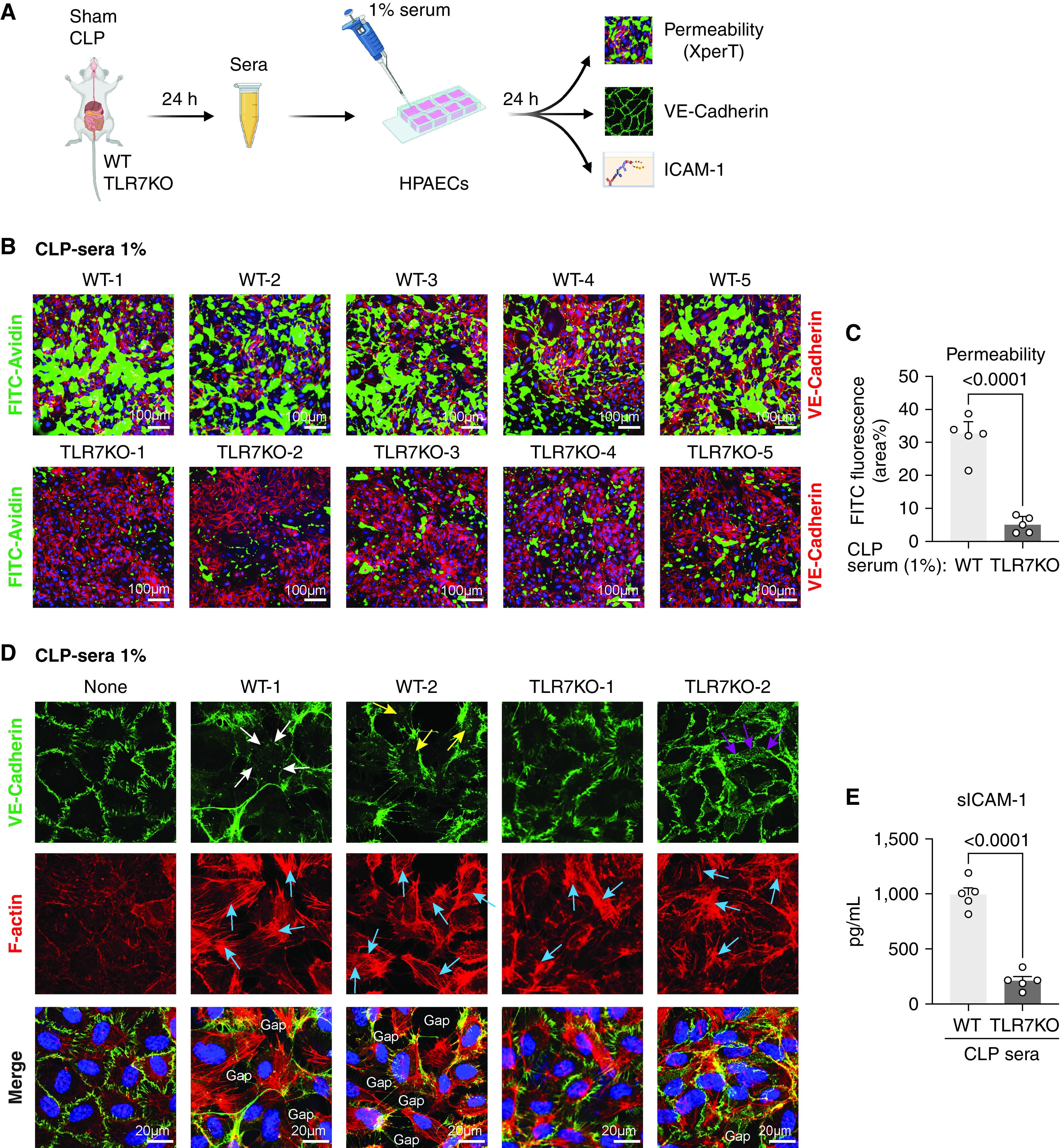
The absence of TLR7 prevents EC barrier disruption by septic sera. (A) Schematic experimental flowchart. ECs were treated with 1% sera collected 24 hours after sham or CLP surgery in WT or TLR7−/− mice. After 24 hours of incubation, permeability was measured by XperT assay, VE-cadherin and F-actin were stained by immunofluorescence, and media ICAM-1 was tested by ELISA. (B–C) Endothelial permeability visualized by XperT and quantified by the area percentage of FITC. n = 5 per group. (D) VE-cadherin and stress fiber formation in ECs treated with 1% sera. White arrowheads: disrupted VE-cadherin; yellow arrowheads: internalized VE-cadherin; purple arrowheads: VE-cadherin network; blue arrowheads: stress fiber. (E) sICAM-1 in the media from treated ECs. n = 5 per group. Data were presented by mean ± SEM and analyzed by unpaired t test.
