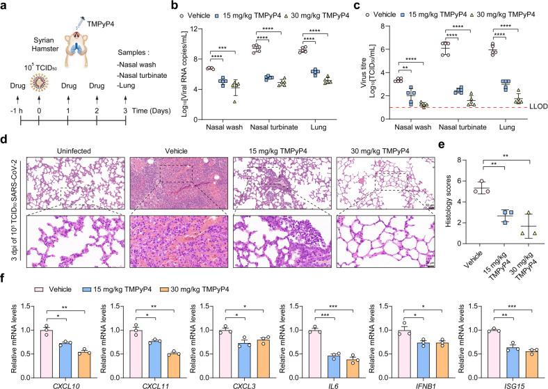
Fig. 6. Antiviral activity of TMPyP4 in SARS-CoV-2-infected hamster.
a Schematic of SARS-CoV-2 infected hamster model. Hamsters infected with SARS-CoV-2 (105 TCID50 virus/hamster) were treated with vehicle (n = 5), 15 mg/kg (n = 5) or 30 mg/kg (n = 5) TMPyP4 for consecutive 3 days, with the first dose given at 1 h before infection with SARS-CoV-2. b, c Viral RNA copies (b) and viral titers (c) in the hamster nasal wash, nasal turbinate and lung tissues of the TMPyP4-treated groups relative to vehicle controls, determined by qRT-PCR and TCID50 at day 3 after infection. LLOD for viral titers is indicated with a red dotted line. d The representative images of hamster lung histopathological changes at day 3 after infection. The corresponding higher-magnification images were shown. See Supplementary Fig. S20 for whole-lung tissue scan images of all hamsters. e Pathological severity scores in hamsters with SARS-CoV-2 infection. f Representative chemokine and cytokine assessment of the lung tissues (n = 3) of the indicated groups, as detected in lung tissue homogenate at day 3 after infection. Data are shown as means ± SEM, two-tailed Student’s t-test. *P < 0.05, **P < 0.01, ***P < 0.001, ****P < 0.0001.

