. 2022 Aug 26;42(7):1897–1911. doi: 10.1148/rg.220045

Figure 11.

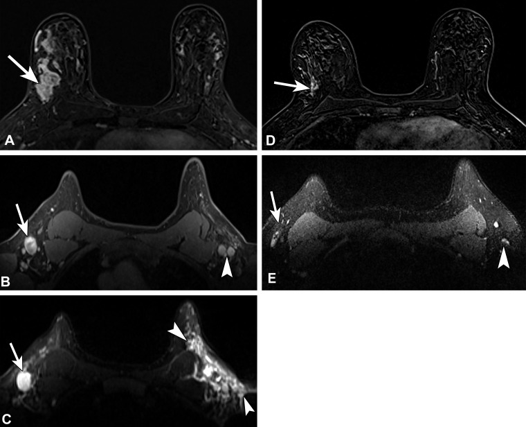
Recently diagnosed invasive ductal carcinoma of the right breast with right axillary nodal metastasis in a 48-year-old woman who presented for MRI for extent of disease evaluation. Three days earlier, she received the first dose of the Pfizer-BioNTech SARS-CoV-2 vaccine in the left arm. (A) Axial contrast-enhanced subtraction MR image shows the known malignancy (arrow) in the right breast. (B, C) Pretreatment axial MR images through the axilla show the known right axillary nodal metastasis (arrow) and left axillary lymphadenopathy (arrowhead in B). On a T2-weighted image (C), there is substantial edema (arrowheads) throughout the left axilla and axillary tail region. US-guided biopsy of an enlarged left axillary lymph node yielded reactive changes. (D) Three-month follow-up axial MR image after neoadjuvant chemotherapy shows decreased extent of disease (arrow). (E) Posttreatment axial MR image through the axilla shows decreased right axillary lymphadenopathy (arrow) and normalized left axillary lymph nodes (arrowhead), as well as resolution of the axillary edema.

Recently diagnosed invasive ductal carcinoma of the right breast with right axillary nodal metastasis in a 48-year-old woman who presented for MRI for extent of disease evaluation. Three days earlier, she received the first dose of the Pfizer-BioNTech SARS-CoV-2 vaccine in the left arm. (A) Axial contrast-enhanced subtraction MR image shows the known malignancy (arrow) in the right breast. (B, C) Pretreatment axial MR images through the axilla show the known right axillary nodal metastasis (arrow) and left axillary lymphadenopathy (arrowhead in B). On a T2-weighted image (C), there is substantial edema (arrowheads) throughout the left axilla and axillary tail region. US-guided biopsy of an enlarged left axillary lymph node yielded reactive changes. (D) Three-month follow-up axial MR image after neoadjuvant chemotherapy shows decreased extent of disease (arrow). (E) Posttreatment axial MR image through the axilla shows decreased right axillary lymphadenopathy (arrow) and normalized left axillary lymph nodes (arrowhead), as well as resolution of the axillary edema.