. 2022 Aug 26;42(7):1897–1911. doi: 10.1148/rg.220045

Figure 12.

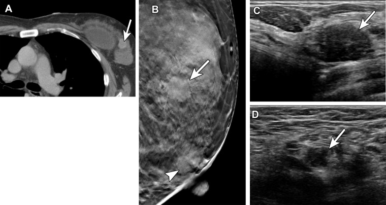
Metastatic carcinoma in a 53-year-old woman with a suspicious mass in the left breast at staging CT, performed after spinal biopsy yielded pathologic findings suggestive of a breast primary. She received the first dose of the Pfizer-BioNTech SARS-CoV-2 vaccine in the left arm 2 days before diagnostic breast imaging. (A) Axial image from staging CT shows the suspicious mass (arrow) in the upper outer left breast. (B) MLO tomosynthesis image shows the corresponding irregular mass (arrow) in the superior left breast. An oval mass (arrowhead) in the subareolar left breast was stable when compared with prior imaging (not shown). (C) US image shows a 1.7-cm mass with irregular margins (arrow) in the left breast at the 2-o’clock position. US-guided core needle biopsy yielded invasive ductal carcinoma. (D) US image of the left axilla shows a corresponding type 6 lymph node (arrow) (BI-RADS 5). Fine-needle aspiration yielded single and clusters of atypical epithelial cells, indicative of metastatic carcinoma.

Metastatic carcinoma in a 53-year-old woman with a suspicious mass in the left breast at staging CT, performed after spinal biopsy yielded pathologic findings suggestive of a breast primary. She received the first dose of the Pfizer-BioNTech SARS-CoV-2 vaccine in the left arm 2 days before diagnostic breast imaging. (A) Axial image from staging CT shows the suspicious mass (arrow) in the upper outer left breast. (B) MLO tomosynthesis image shows the corresponding irregular mass (arrow) in the superior left breast. An oval mass (arrowhead) in the subareolar left breast was stable when compared with prior imaging (not shown). (C) US image shows a 1.7-cm mass with irregular margins (arrow) in the left breast at the 2-o’clock position. US-guided core needle biopsy yielded invasive ductal carcinoma. (D) US image of the left axilla shows a corresponding type 6 lymph node (arrow) (BI-RADS 5). Fine-needle aspiration yielded single and clusters of atypical epithelial cells, indicative of metastatic carcinoma.