. 2022 Aug 26;42(7):1897–1911. doi: 10.1148/rg.220045

Figure 13.

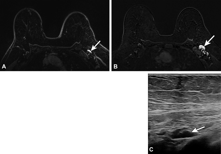
Type 5 lymph node in a 62-year-old woman with human epidermal growth factor receptor 2 (HER2)–positive invasive ductal carcinoma of the left breast, who presented for MRI evaluation of response to neoadjuvant chemotherapy. Three weeks earlier, she received the third (booster) dose of the Moderna SARS-CoV-2 vaccine in the left arm. (A) Pretreatment axial MR image shows a normal-appearing level 1 lymph node (arrow). (B) Posttreatment axial MR image shows enlargement of the lymph node (arrow). (C) Same-day US image of the left axilla shows a type 5 lymph node (arrow) (BI-RADS 4). US-guided core biopsy yielded lymphoid tissue with lymphocytes and histiocytes, without evidence of metastatic carcinoma.

Type 5 lymph node in a 62-year-old woman with human epidermal growth factor receptor 2 (HER2)–positive invasive ductal carcinoma of the left breast, who presented for MRI evaluation of response to neoadjuvant chemotherapy. Three weeks earlier, she received the third (booster) dose of the Moderna SARS-CoV-2 vaccine in the left arm. (A) Pretreatment axial MR image shows a normal-appearing level 1 lymph node (arrow). (B) Posttreatment axial MR image shows enlargement of the lymph node (arrow). (C) Same-day US image of the left axilla shows a type 5 lymph node (arrow) (BI-RADS 4). US-guided core biopsy yielded lymphoid tissue with lymphocytes and histiocytes, without evidence of metastatic carcinoma.