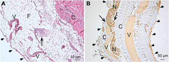
FIGURE 1.

Light microscopic findings. A, H&E staining of a longitudinal section of a filum specimen. A pial cell layer envelopes the FT (arrowheads). Underneath a larger fat pad (F), the collagenous core of the FT can be seen. The fat contains multiple small vessels (V) and, in just 1 specimen, a Pacinian mechanoreceptor (arrow). Pacinian corpuscles are typically found in subcutaneous tissue of the skin and are pressure sensitive.12 B, Immunostaining of a longitudinal section of a filum specimen. The arrowheads mark the outer pial cell layer. A nerve (N) is piercing through the pia into the fibrous core of the FT (arrows). Multiple blood vessels (V) are running in a longitudinal direction within the collagenous tissue C. FT, filum terminale.
