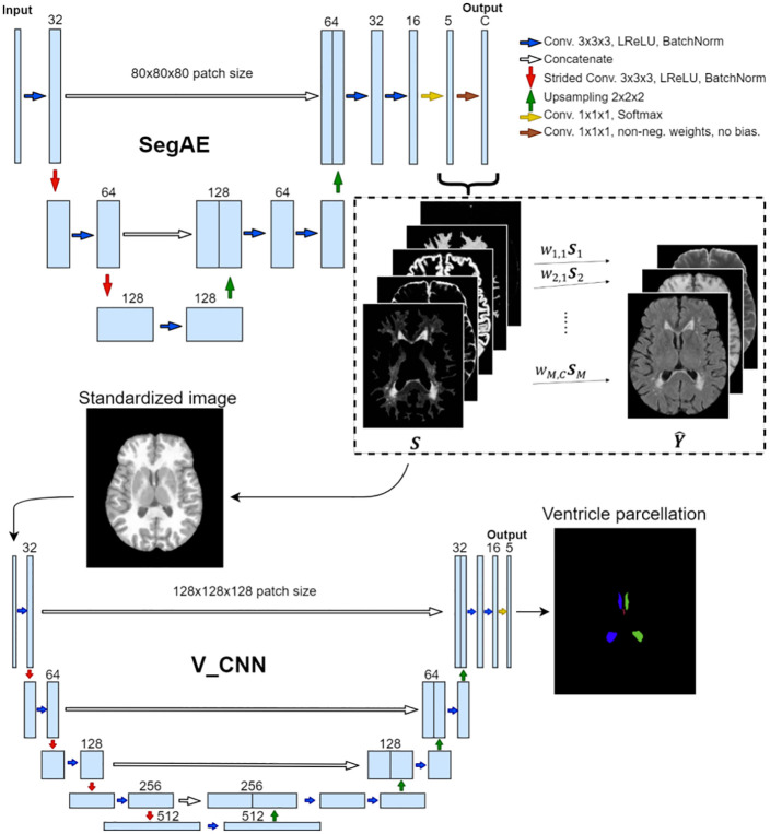
Fig 5. The proposed CNN architecture.
The input into the SegAE network comprises large 3D patches of MRI images that are in turn reconstructed in an unsupervised manner by SegAE (the reconstructed output is denoted with ). The estimated components of the reconstruction (denoted with S) provide the segmentation of the input into WMHs, WM, GM, CSF, and meninges (meninges are discarded in subsequent steps), which in turn are used to create a standardized image that is used as the input into the V_CNN. Kernels of size 3 × 3 × 3 are used in all convolutional layers except size 1 × 1 × 1 is used in the final two layers of both SegAE and the V_CNN. The V_CNN output is a segmentation of the four ventricle compartments, which in conjunction with the SegAE output provides a consistent ventricle and WMH segmentation.

