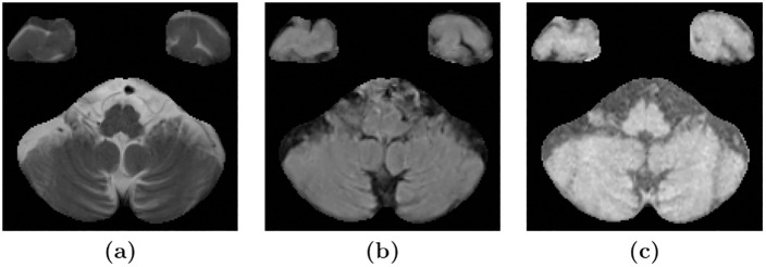
Fig 10. Resampling may cause erroneous WMH segmentation in the cerebellum.
Images (a), (b), and (c) show an axial slice of the cerebellum in T2-w, FLAIR, and T1-w images, respectively, of a subject in the NPH data set. The T2-w images have a higher in-plane resolution, which shows the thin lines of CSF in the cerebellum. Meanwhile, the upsampling of the lower resolution FLAIR and T1-w images gives them a blurry appearance, leading to brighter voxels instead of fine dark lines corresponding to the CSF in the T2-w image.

