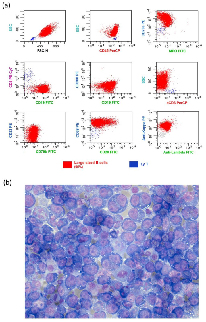
Figure 1.
Flow cytometric immunophenotype and cytological examination of ascitic fluid. (a) Flow cytometric immunophenotype of neoplastic cells from ascitic fluid (95% of infiltration). The pathological cells appear as highly scattered lymphoid cells (colored red) positive for CD45/CD79a/CD22/CD19/CD38 partially expressing CD20 (20%), kappa light chain restricted and negative for CD5/CD200/CD79b/cCD3. Colored blue normal T lymphocytes; (b) Cytological examination of ascitic fluid with May-Grunwald-Giemsa stain. Cytologic preparations appear highly cellular and composed of lymphoid cells of medium/large size often in apoptosis. Lymphoma cells are characterized by round nuclei, prominent nucleoli and abundant deeply basophilic and microvacuolated cytoplasm and show occasional mitotic figures. Several binucleated cells are also present.

