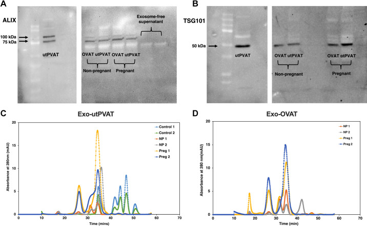
Figure 2.
Characterization of exosome-related protein markers and molecular masses in vesicles isolated from adipose-tissue culture media. Protein content of Alix (100 and 75 kDa; A) and TSG101 (∼50 kDa; B) in samples of Exo-utPVAT and Exo-OVAT derived from nonpregnant and pregnant rats. Representative chromatograms fast protein liquid chromatography (FPLC) for Exo-utPVAT (C) and Exo-OVAT samples (D), demonstrating time at which the protein eludes in the FPLC column. Exo-OVAT, exosome-like extracellular vesicles derived from periovarian adipose tissue; Exo-utPVAT, exosome-like extracellular vesicles derived from uterine perivascular adipose tissue; utPVAT, uterine perivascular adipose tissue.

