Figure 1.
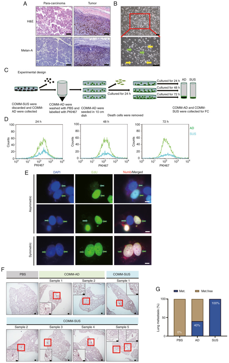
Malignant mucosal melanoma cells exhibited a marked heterogeneous phenotype and biological behaviors. (A) H&E and immunohistochemistry of COMM specimens. Melan-A indicated COMM cells. Scale bar, 100.0 µm. (B) Two subpopulations of COMM cells are indicated by the arrows; green arrows indicate COMM-AD cells (abbreviated as AD), and yellow arrows indicate COMM-SUS cells (abbreviated as SUS). (C) Schematic diagram of the experimental design of flow cytometric analysis. COMM-SUS cells were discarded and the whole adherent cell population was labeled with PKH67, and the cells were then seeded into culture plates for 24 h. After rinsing with the same medium, the cells were further cultured for 24, 48 and 72 h, and then analyzed by flow cytometry. (D) The results of flow cytometry of PKH67-labeled cells. (E) Immunofluorescence of template DNA co-segregate asymmetric and symmetric division. EdU (green) staining indicated distribution of DNA. Positive EdU (yellow arrows) indicated the DNA from parent cells. Negative EdU (white arrows) indicated the newly synthesized DNA. DAPI (blue) staining indicated nuclei. Numb (red) staining indicated the asymmetric division of the cell membrane. Scale bar, 10.0 µm. (F) Lung metastases were analyzed after tail intravenous injections of COMM-AD and COMM-SUS cells with eGFP, respectively at 4 weeks. Positive melan-A staining indicated COMM cells in lungs. Scale bar, 100.0 µm. (G) The frequency of developed lung metastasis in mice, expressed as a percentage (Met, metastasis-positive; Met.free, metastasis-free). n=5 mice per group. H&E, hematoxylin and eosin; COMM, Chinese oral mucosal melanoma; COMM-AD, cells with adhesive morphology; COMM-SUS, cells grown in suspension.

