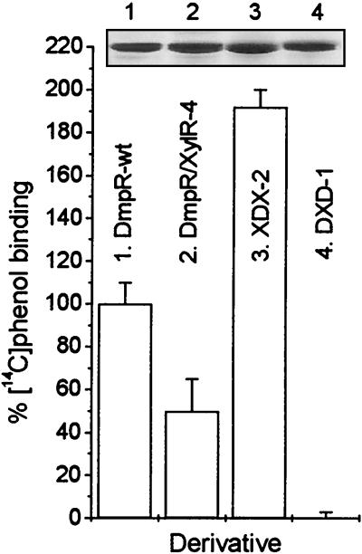
FIG. 5.
Comparison of [14C]phenol binding of DmpR-Flag and DNA-shuffling derivatives. The data are the averages (+ standard deviations) of triplicate determinations performed at the Kd for wild-type DmpR-Flag (16 μM) as described in Materials and Methods. Binding by wild-type DmpR-Flag was set at 100%. The inset shows a Coomassie blue stain of 3 μg of proteins released from 3 μl of the beads and separated by SDS–11% PAGE.

