. 2022 Sep 6;11:e78416. doi: 10.7554/eLife.78416

Figure 4. Electrophysiological measurements of related proteins of viral HeR from Emiliania huxleyi virus 202 (V2HeR3) and a functional model.

(A) Electrophysiological measurements of a heliorhodopsin (HeR) from the host Emiliania huxleyi (Ehux), two HeRs from E. huxleyi virus 201 (V1HeR1, 2) and three HeRs from E. huxleyi virus 202 (V2HeR1-3). Although some HeRs exhibit transient photocurrents (positive or negative peaks), steady-state photocurrent was only observed for V2HeR3. Comparison of photocurrent densities at −40mV. Square-block bar indicates amplitude from peak photocurrent (I1), open bar indicates amplitude from steady-state photocurrent (I2). The pipette solution was 110mM NaCli, pH 7.4i, the bath solution was 140mM NaCle, pH 7.4e. n=5–8cells. (B) Electrophysiological measurements and the obtained I-V plots of homologous proteins of V2HeR3. Comparison of photocurrent amplitudes at −40mV. Square-block bar indicates amplitude from peak photocurrent (I1), open bar indicates amplitude from steady-state photocurrent (I2). The pipette solution was 110mM NaCli, pH 7.4i, the bath solution was 140mM NaCle, pH 7.4e. n=5–8cells. (C) Key residues for ion transport of HeRs. (D) Schematic drawing of suggested proton-transporting mechanism in V2HeR3.

Figure 4.

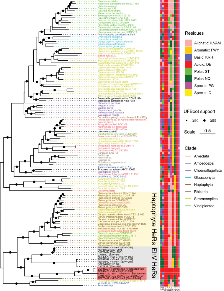
Figure 4—figure supplement 1. Phylogenetic relationships of the EhV heliorhodopsins (HeRs) and their eukaryotic cognates.

Figure 4—figure supplement 1.

Eukaryotic HeRs most similar to HeRs from EhV were collected from a collection of transcriptomes and genomes of algae and other unicellular eukaryotes. Eukaryotic HeRs are named after the corresponding species/strains and colored by their taxonomic origin. The tree is midpoint-rooted which places HeRs from EhVs as a sister clade to the predominantly haptophyte HeRs, a group that also includes EhHeR from the host species, Emiliania huxleyi. Distribution of the amino acid residues among a selected set of viral HeR from Emiliania huxleyi virus 202 (V2HeR3) positions is indicated to the right. Data and code used to generate the phylogeny are available from https://github.com/BejaLab/ViralHeRs, (copy archived at swh:1:rev:0a2cc9a502c5d0d9dbc6dbcac962d89c3a5a254d, Rozenberg, 2022).

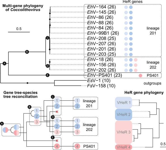
Figure 4—figure supplement 2. Evolution of coccolithoviruses and their heliorhodopsin (HeR) genes.

Figure 4—figure supplement 2.

Multigene phylogeny of EhV isolates using 26 single-copy genes (number of orthogroups used per genome is indicated in parentheses) was rooted using two genomes from of phaeoviruses (top). Black dots represent highly supported branches (ultrafast bootstrap support≥95). For each EhV genome the presence of HeR genes from the four HeR clades is indicated with circles colored according to the HeR phylogeny (bottom right). The two clades showing proton transport are highlighted in red. Gene tree-species tree reconciliation providing a hypothesis about the evolution of the HeR genes among EhVs (bottom left) is based on the assumption of no horizontal transfer of HeR genes between viruses. ⊏ forks represent co-divergence, ⊂ forks represent gene duplication events, × representsgene losses. The numbers in the black circles correspond to branches on the species tree. List of phylogenetic markers, accession numbers of HeR genes, and lineage assignments are available in Supplementary file 2. Code used to generate the phylogenies is available from https://github.com/BejaLab/ViralHeRs.

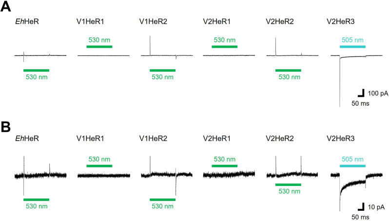
Figure 4—figure supplement 3. Representative photocurrent traces of heliorhodopsins (HeRs) from Emiliania huxleyi and its viruses.

Figure 4—figure supplement 3.

(A) Electrophysiological measurements of virus HeRs-driven photocurrent in ND7/23cells. The cells were illuminated with light (λ=505nm, 24.5 mW/mm2, λ=530nm, 27.8 mW/mm2) during the time region shown by green bars. The membrane voltage was clamped at –40mV. The pipette solution was 110mM NaCli, pH 7.4i, the bath solution was 140mM NaCle, pH 7.4e. (B) The same traces as (A) with expanded Y axis.

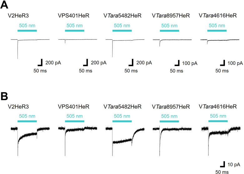
Figure 4—figure supplement 4. Representative photocurrent traces of viral HeR from Emiliania huxleyi virus 202 (V2HeR3)-like heliorhodopsins (HeRs).

Figure 4—figure supplement 4.

(A) Electrophysiological measurements of V2HeR3-like HeRs-driven photocurrent in ND7/23cells. The cells were illuminated with light (λ=505nm, 24.5 mW/mm2) during the time region shown by blue bars. The membrane voltage was clamped at –40mV. The pipette solution was 110mM NaCli, pH 7.4i, the bath solution was 140mM NaCle, pH 7.4e. (B) The same traces as (A) with expanded Y axis.

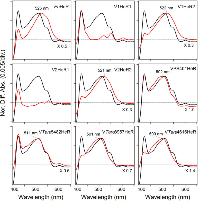
Figure 4—figure supplement 5. Absorption spectra of heliorhodopsins (HeRs) from Emiliania huxleyi and its viruses, and viral HeR from Emiliania huxleyi virus 202 (V2HeR3)-like HeRs obtained by the hydroxylamine bleach of ND7/23cells.

Figure 4—figure supplement 5.

Light-induced difference absorption spectra in the presence of 50mM hydroxylamine of HeRs from E. huxleyi and its viruses, and V2HeR3-like HeRs (red line). Absorption spectra were normalized by use of a positive peak at each λmax.

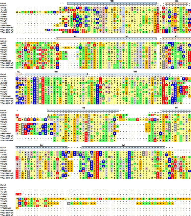
Figure 4—figure supplement 6. Alignment of heliorhodopsins (HeRs).

Figure 4—figure supplement 6.

Multiple amino acid alignment of viral HeR from Emiliania huxleyi virus 202 (V2HeR3) with 48C12, TaHeR, EhHeR, V1HeR1, V1HeR2, V2HeR1, V2HeR2, VPS401HeR, VTara5482HeR, VTara4616HeR, and VTara8957HeR.