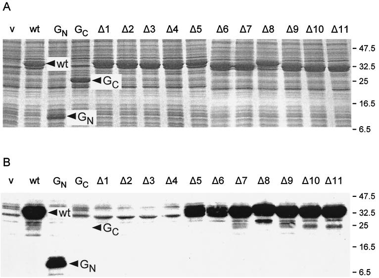
FIG. 4.
(A) Coomassie blue-stained gels of whole cell protein from E. coli BL21(DE3)pLysS transformed with pET-based plasmids expressing various alleles of fliG and grown in the presence of IPTG. (B) Affinity blot of the same samples using purified N-His-FLAG-FliF as a probe. v (vector), pET19b; wt, pGMK3000 (wild-type FliG); GN, pGMK3100 (N-terminal fragment of FliG, residues 1 to 108); GC, pGMK3200 (C-terminal fragment of FliG, residues 109 to 331); Δ1, etc., pGETΔ1, etc. Molecular mass markers are indicated on the right in kilodaltons.

