Figure 1.

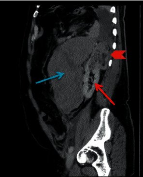
Contrast sagittal section shows a large hematoma at the left retroperitoneal region (blue arrow), the normal-appearing left kidney (thin red arrow), and an exophytic left upper pole renal mass (red arrowhead).

Contrast sagittal section shows a large hematoma at the left retroperitoneal region (blue arrow), the normal-appearing left kidney (thin red arrow), and an exophytic left upper pole renal mass (red arrowhead).