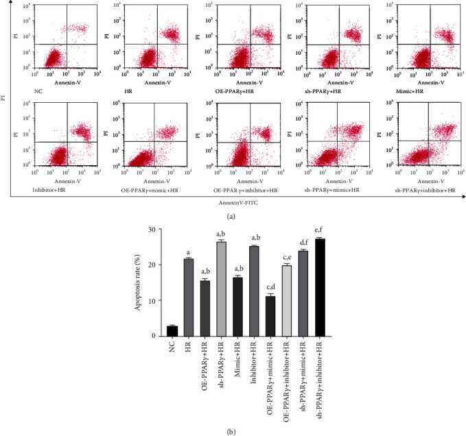
Figure 3.

Flow cytometric analysis was used to examine cell apoptosis (n = 4): (a) representative images of flow cytometric analysis; (b) quantitative analysis of apoptosis rate (A represents values compared to the NC group, p < 0.05; B represents values compared to the HR group, p < 0.05; C represents values compared to the OE-PPARγ+HR group, p < 0.05; D represents values compared to the mimic+HR group, p < 0.05; E represents values compared to the inhibitor+HR group, p < 0.05; F represents values compared to the sh-PPARγ+HR group, p < 0.05).
