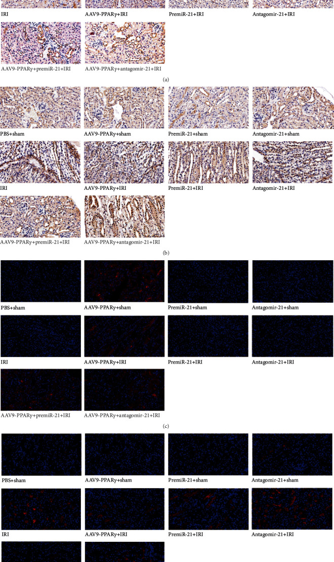
Figure 8.

Immunohistochemistry and immunofluorescence analysis in the kidney (n = 5): (a) immunohistochemistry analysis of PPARγ; (b) immunohistochemistry analysis of PDCD4 (light-yellow or brown color cells were regarded as positive); (c) immunofluorescence analysis of PPARγ; (d) immunofluorescence analysis of PDCD4 (red was the positive part, all scale bar 20 μm).
