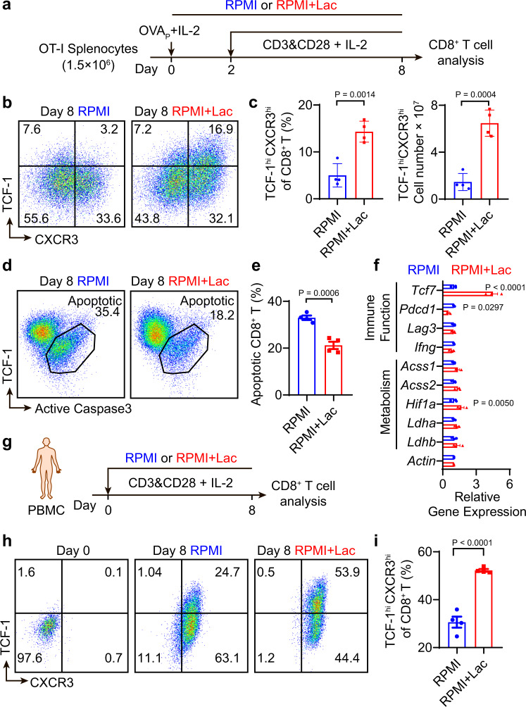
Fig. 5. Lactate increases TCF-1 expression and reduces apoptosis of CD8+ T cells during ex vivo expansion.
a Experimental design of ex vivo OT-I CD8+ T cell expansion. Fresh splenocytes from C57BL/6-Tg(TcraTcrb)1100Mjb/J mice were primed with SIINFEKL peptide (1 μg/mL) and hIL-2 (50 U/mL) for two days and stimulated with anti-CD3 and anti-CD28 (0.5 μg/mL each) from day 3 to 8 with hIL-2 (30 U/mL). b, c Flow cytometry plots and quantification of TCF-1hiCXCR3hi population of OT-I CD8+ T cells on day 8 of ex vivo expansion (n = 4 biologically independent samples). d, e Quantification of percentage of apoptotic OT-I CD8+ T cells by flow cytometry on day 8 of ex vivo expansion (n = 4 biologically independent samples). f, Relative gene expression in OT-I CD8+ T cells on day 4 detected by RT-PCR (n = 3 biologically independent samples). g Experimental design of ex vivo expansion of CD8+ T cells from human PBMCs. PBMCs from cord blood were activated and cultured in the presence of anti-CD3 and anti-CD28 beads (T cell: Beads = 1: 1) supplemented with hIL-2 (30 U/mL). h, i Flow cytometry plots and quantification of TCF-1hiCXCR3hi population of human CD8+ T cells on day 8 of ex vivo expansion (n = 4 biologically independent samples). Data are shown as means ± SEM. P-value was determined by two-tail unpaired t-test (c, e, i) or one-tail two-way ANOVA without correction (f). Source data are provided in Source Data file.

