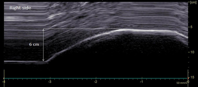
FIGURE 2.
Diaphragmatic motion recorded by M-mode ultrasonography in a man suffering from left hemidiaphragm dysfunction. Hemidiaphragm excursions were measured by placing the first caliper at the foot of the inspiration slope on the diaphragmatic echoic line and by placing the second caliper at the apex of the curve (see arrow). On the right side: normal excursion during deep breathing (6 cm for a lower limit of normal = 4.1 cm).

