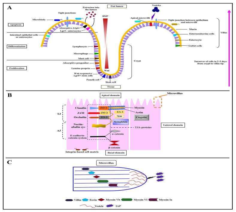
Figure 1.
A depiction of epithelial cell polarity. (A) Turnover of cells in 2–5 days from crypt to villus tip and (B) α-catenin in Figure 1. (C) The box area from panel B describes the single microvillus within its protein components such as villin, ezrin, myosin Vb, myosin-VI, and myosin-Ia, and the microvillus-derived vesicle with alkaline phosphatase enzyme.

