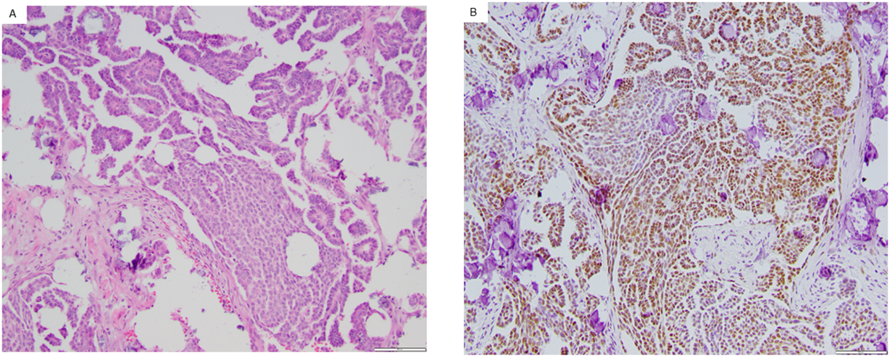
Figure 1:

Representative example of an Androgen Receptor positive low-grade serous ovarian carcinoma. Androgen Receptor (AR) expression was assessed in archival (fresh frozen paraffin embedded) or fresh tissue. Pictured is the hematoxylin and eosin stain of a low-grade serous carcinoma (A) as well as AR+ immunohistochemistry (B). Both images obtained with magnification of 100x.
