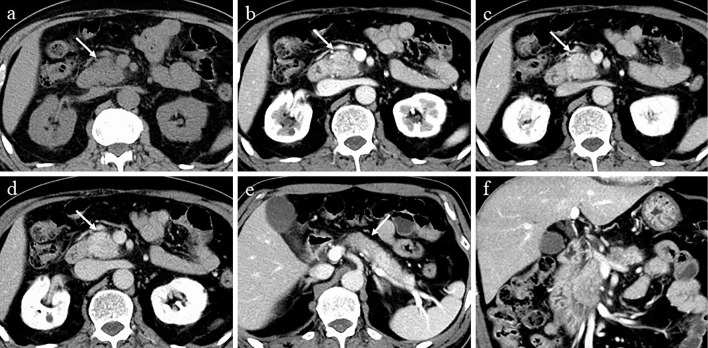
Figure 1.
CT at the time of the diagnosis. a-d: CT showing localized enlargement and delayed enhancement in the head of the pancreas. a: Unenhanced phase, b: parenchyma phase, c: portal phase, d: equilibrium phase. e: CT showing hypoattenuating areas in the body of the pancreas in the parenchyma phase (arrow). f: Coronal section in the parenchyma phase. CT: computed tomography

