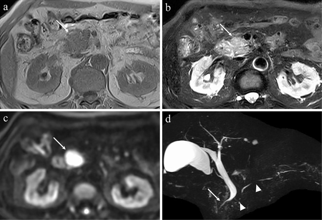
Figure 3.
MRI findings. MRI showing enlargement of the head of pancreas (arrows) with a low signal on T1-weighted imaging (a), faint high signal on T2-weighted imaging (b), and strong high signal on diffusion-weighted imaging (c). Magnetic resonance cholangiopancreatography showing multiple stenoses in the main pancreatic duct (arrowheads) and stenosis of the intrapancreatic bile duct (d) (arrow). MRI: magnetic resonance imaging

