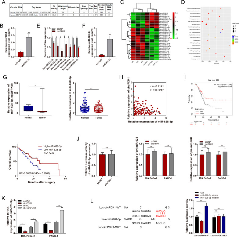
Fig. 4.
circPDK1 functions as a miR-628-3p sponge in PC. A The potential binding site between circPDK1 and AGO2 were predicted by circinteractome. B RIP assay was used to determine the relative expression of circPDK1 with rabbit AGO2 and IgG antibodies in MIA PaCa-2 cells. C Cluster heatmap showing the differentially expressed miRNAs between control and circPDK1 overexpression in MIA PaCa cells. D Pathway enrichment analysis were analyzed by using differentially expressed miRNAs after circPDK1 overexpression. E Seven potential target miRNAs expression in MIA PaCa-2 after circPDK1 overexpression. F RIP assay was used to determine the relative expression of miR-628-3p with rabbit AGO2 and IgG antibodies in MIA PaCa-2 cells. G The relative expression of miR-628-3p in PC patients were detected by the data from TCGA and our center. H The correction between circPDK1 and miR-628-3p were determined from our center. I Prognosis analysis of miR-628-3p were detected using survival data of PC patients from TCGA and our center. J Promoter luciferase activity in 293 T cells, pri-miR-628-3p and pre-miR-628-3p expression were verified after circPDK1 overexpression. K The expression of miR-628-3p were detected after circPDK1 overexpression and loss of circPDK1 in MIA PaCa-2 and PANC-1 cells. L Luciferase activity in 293-T cells co-transfected with Luc-circPDK1 wild-type or mutant sequence and miR-628-3p mimics or inhibitor. *P < 0.05; **P < 0.01; ***P < 0.001; ns, no significance

