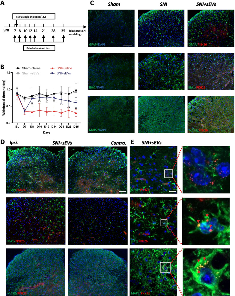
Fig. 2.
Analgesic effect of hPMSCs sEVs and localization in spinal cord injected intrathecally in mice. A Experimental schematic plot for the establishment of the mouse model of spared nerve injury (SNI). B 50% paw withdraw threshold (PWT) of left hind paw of different treatment groups mice. At the day of 8, 10, 12, 14, 21, 28 and 35, the paw withdrawal thresholds (WTs) of SNI + sEVs group was significantly higher than those of SNI + saline group after single intrathecal injection at the 7th day (**p < 0.01 compared with SNI + saline group by Two-way ANOVA followed by Tukey post hoc test, n = 8 in each group). C Immunofluorescence study showed GFAP astrocyte/IBA1 microglia/MAP2 neurons (green) in the dorsal horn of the L4–5 spinal cord of SNI mice and changes after intrathecal injection of sEVs (PKH26). The blue spots are DAPI nuclear staining. Scale bar: 50 μm. D Immunofluorescent study revealed pkh26 labeled hPMSC sEVs (red) localized within GFAP astrocyte/IBA1 microglia/MAP2 neuron (green) in the L4–5 spinal dorsal horn of SNI mice. The blue spots are DAPI nuclear staining (Scale bar: 100 μm). E pkh26 labelled hPMSCs sEVs appeared majorly the in the ipsilateral (Ipsi.) L4–5 spinal dorsal horn (DH) of SNI mice and co-localized mostly with MAP2 neuron. Some sEVs appeared in the IBA1 microglia. Few sEVs could be detected in the GFAP astrocyte. The blue spots are DAPI nuclear staining (Scale bar: 20 μm)

