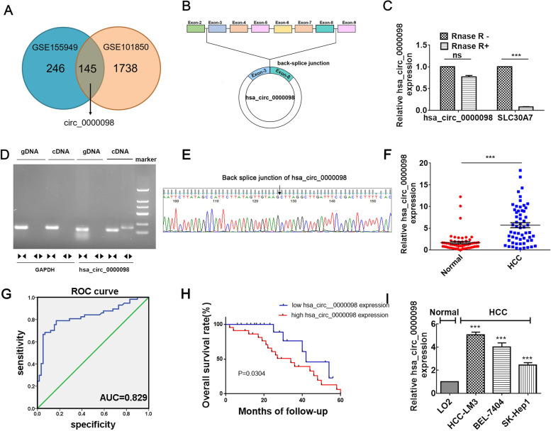
Fig. 1.
Circ_0000098 is highly expressed in HCC. A Venn diagrams from analysis of GSE155949 and GSE101850 datasets. B Illustration of circ_0000098 formation from SLC30A7 gene. C The stability of circ_0000098 and SLC30A7 mRNA was evaluated following RNase R treatment using RT-qPCR assays. D Assay of PCR-amplified products of circ_0000098 on an agarose gel electrophoresis. Divergent primers were capable of amplifying circ_0000098 inside cDNA but not genomic DNA (gDNA). GAPDH served as a linear reference, while convergent primers amplified both circ_0000098 and linear gDNA. E The back-splicing junction of circ_0000098 was validated using Sanger sequencing. F Comparison of circ_0000098 levels in HCC and adjacent normal tissues. G ROC curve analysis of circ_0000098 for distinguishing HCC from healthy tissues. H Kaplan–Meier curve analysis of the prognosis of HCC patients with high or low circ_0000098 expression. I RT-qPCR was used to detect circ_0000098 expression in HCC cells (HCC-LM3, BEL-7404, and SK-Hep1) and a normal cell line LO2. NS: not significant. ***P < 0.001

