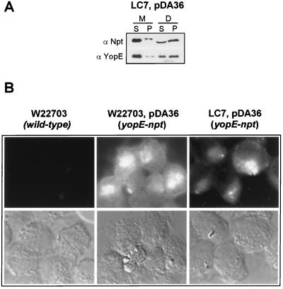
FIG. 3.
Y. enterocolitica LC7 (tyeA2) targets YopE-Npt into the cytosol of HeLa cells. (A) HeLa cell cultures were infected with Y. enterocolitica LC7 (tyeA2) carrying pDA36, expressing the YopE-Npt fusion protein (23), fractionated by the digitonin technique (see the legend to Fig. 2), and analyzed by immunoblotting. α-Npt measures the distribution of YopE-Npt in various fractions, whereas α-YopE measures the distribution of YopE. (B) Immunofluorescence microscopy of HeLa cells infected with Y. enterocolitica W22703, W22703(pDA36), or LC7(pDA36). Samples were fixed with formaldehyde and stained with α-Npt antibodies followed with an α-rabbit IgG-FITC conjugate. YopE-Npt staining was detected in the cytosol of HeLa cells infected with W22703(pDA36) and LC7(pDA36) but not in HeLa cells that were infected with strain W22703.

