
Figure 1.
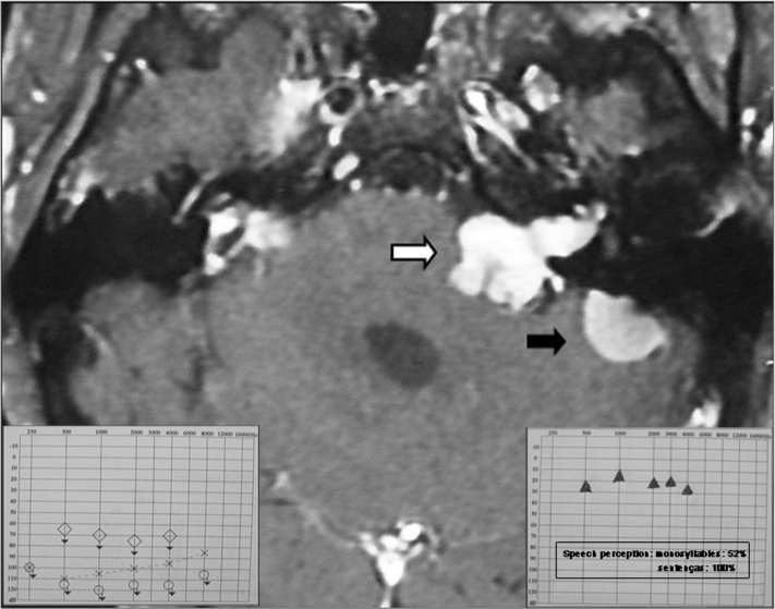
Magnetic resonance imaging (T1 contrast) showing a vestibular schwannoma to the left (white arrow) associated with a meningioma on the medial aspect of the left petrous bone (black arrow). Note also small manipulation remains in the right pontocerebellar angle. On the lower left detail, see preoperative audiometry. On the lower right detail see audiometry 6 months after the cochlear implant.