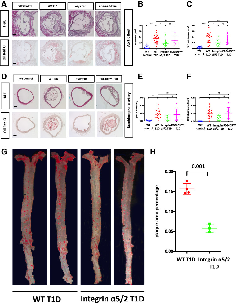
Figure 2.
Atherosclerosis in arteries from mice injected with PCSK9 virus and placed on a HFD for 8 weeks. (A) Representative images of H&E and ORO staining of aortic root sections. (B) Quantification of plaque area of H&E staining in A. (C) Quantification of aortic root ORO staining area in A. (D). H&E and ORO staining of brachiocephalic artery (BCA) cross sections. (E) Quantification of BCA plaque area of H&E staining in D. (F) Quantification of BCA ORO-positive area in D. (G) Representative images of en face preparations of WT T1D and integrin α5/2 T1D aortas stained with ORO. (H) Quantification of ORO-positive area to whole area ratio in G. Analysis was by Student t test. (A–F) WT control, n = 7 mice. WT T1D, n = 16 mice; integrin α5/2 T1D, n = 11; and PDE4Dmut T1D, n = 7. (G and H) WT T1D, n = 4 mice; integrin α5/2 T1D, n = 3. (B, C, E, and F) One-way ANOVA was used. Ctrl, control.

