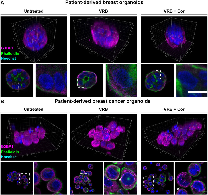
Fig. 3.
VRB and cortisone induce the formation of SGs in patient-derived breast cancer organoids. SG formation was examined when cortisone (Cor; 300 µM) was added with the VRB treatment (75 µM for 2 h) in patient-derived breast organoids originating from either (A) healthy tissue or from (B) cancerous tissue. SGs were detected using an anti-G3BP1 antibody (magenta). Phalloidin staining of actin (green) was used to detect the cell outline. Hoechst 33342 DNA stain is shown in blue. The top rows show 3D presentations of the organoids. Dashed boxes in the images of organoid sections indicate regions of cells shown in enlarged images on the right. Arrowheads mark SGs. Scale bars: 10 µm. Images are representative of three experiments.

