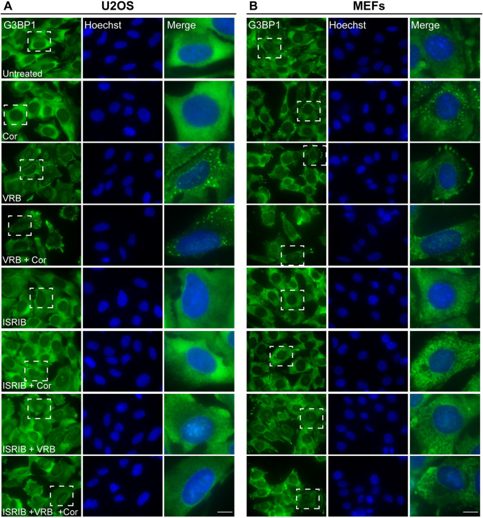
Fig. 7.
ISRIB blocks SG formation induced by VRB and cortisone. (A) U2OS cells and (B) MEFs treated with ISRIB (5 µM) for 2 h before addition of VRB (75 µM for U2OS cells, 15 µM for MEFs), cortisone (Cor, 300 µM) or both for 1 h, as indicated. SGs were detected using G3BP1 (green) as an SG marker. Hoechst 33342 DNA stain is shown in blue. Dashed boxes indicate the regions shown in enlarged images on the right. Scale bars: 10 µm. Images shown are representative of three experiments.

