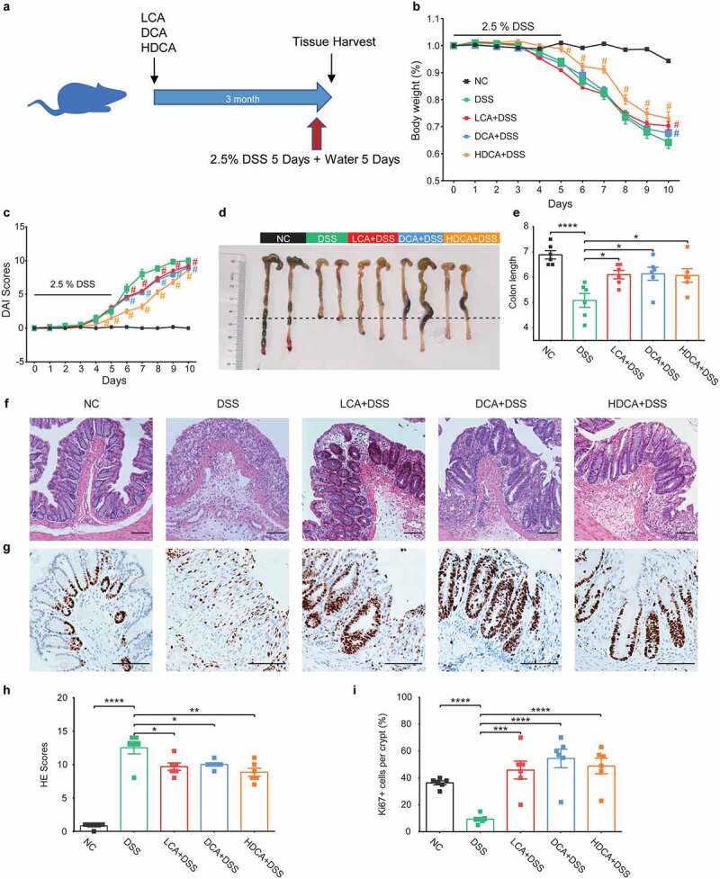
Figure 3.

Secondary BAs also mitigate DSS-induced murine colitis. a. Schematic diagram showing the overall design and complete timeline. Mice were treated with LCA, DCA, or HDCA (2 mM of each) in drinking water for 3 months before inducing colitis. b. Body weight change (relative to starting weight, set as 100%) during the course of DSS-induced colitis. c. Disease activity index during colitis models. d-e. Representative colonic images and colon length. f. Representative hematoxylin and eosin-stained sections of the colon. g. Immunohistochemical staining of Ki67 in colon sections. h. histological scores of colons. i. Quantitative analysis of colonic ki67+ cells. Scale bar, 100 μm. Data are represented as mean ± SEM. N = 6–8 per group. *P < .05, **P < .01, ***P < .001, ****P < .0001, #P < .05 compared with DSS. ns: not significant. BAs, bile acids; DCA, deoxycholic acid; DSS, dextran sulfate sodium; HDCA, hyodeoxycholic acid; HE, hematoxylin & eosin; LCA, lithocholic Acid; NC, normal control.
