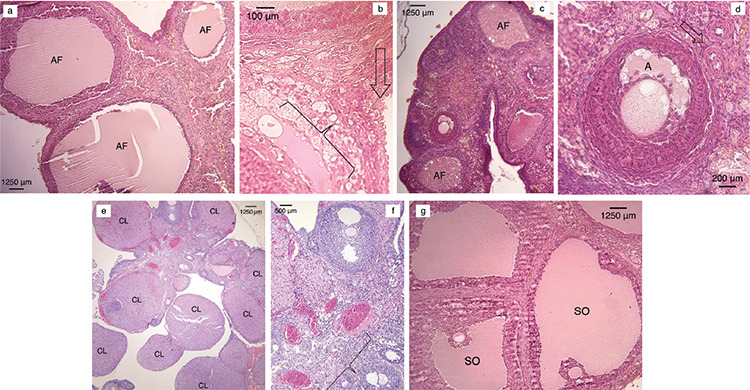
Figure 5.

Photomicrographs of the ovaries of the groups. (haematoxylin & eosin). a) Different stages of degenerated atretic follicles (AF) are evident in the CNT group animal samples (x40 magnification). b) Vacuolation in the interfollicular area (parenthesis) is evident in the CNT group samples in the interstitial area (arrow) (x400 magnification). c) The human umbilical mesenchymal cord stem cells (hUCMSCs)-applied group shows healthy antral follicles (AF) surrounded by a preantral follicle (x40 magnification). d) The hUCMSCs-applied group demonstrating intact zona pellucida and antrum a) and oocytes and primordial follicles (arrow) (x200 magnification). e) The amniotic fluid (AF)-applied group exhibited superovulation and multiple corpus luteum (CL) (x40 magnification). f) The AF-applied group displayed multiple endometrial blood vessels (parenthesis) (x100 magnification). g) Both hUCMSCs and AF-applied groups showed superovulated follicles (SO) (x40 magnification)
