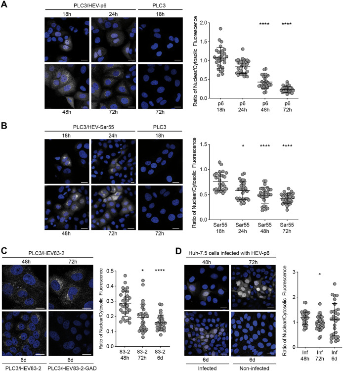
Fig 1. Kinetics of the subcellular localization of ORF2 protein.
PLC3 cells electroporated with HEV-p6 (A), HEV-Sar55 (B) and HEV83-2 (C) RNA and Huh-7.5 cells infected with HEV-p6 (D) were fixed at the indicated timepoints post-electroporation (p.e.) (A-C) or post-infection (p.i.) (D). Indirect immunofluorescence analysis was performed using the 1E6 anti-ORF2 antibody. Cells were analyzed by confocal microscopy (magnification x40). Scale bar, 20 μm. Nuclear/cytosolic fluorescence intensity quantification was done using ImageJ software (mean ± S.D., n ≥ 30 cells, Friedman with Nemenyi test). *p < 0.05, ****p < 0.0001. Mock electroporated PLC3 cells (PLC3), cells electroporated with a replication-deficient HEV83-2 strain (PLC3/HEV83-2-GAD) and non-infected Huh-7.5 cells were used as controls.

